flow cytometry results for lymphoma
Nonhematologic malignancy can be suspected if less than 75 percent of the cells show CD45 common leukocyte antigen. Flow-cytometric demonstration of the typical chronic lymphocytic leukemia CLL immunophenotype is vital for diagnosis.

Flow Cytometry Results Flow Cytometric Graphs Showing Positivity For Download Scientific Diagram
Flow cytometry is rapid and appears to be virtually diagnostic of non-Hodgkins lymphoma when a majority of cells are B cells with an abnormal kappalambda ratio 40 or 025.

. They also expressed the CD38 CD123 CD58 CD81 and HLA-DR. Our results show the feasibility of using flow cytometry to evaluate body fluids or FNA and dem- onstrate that small malignant populations that may be missed by routine cytology. Tumor cells are positive for CD45 the B cell markers CD19 CD22 and CD79a with a monoclonal light chain expression of kappa or lambda.
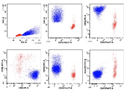
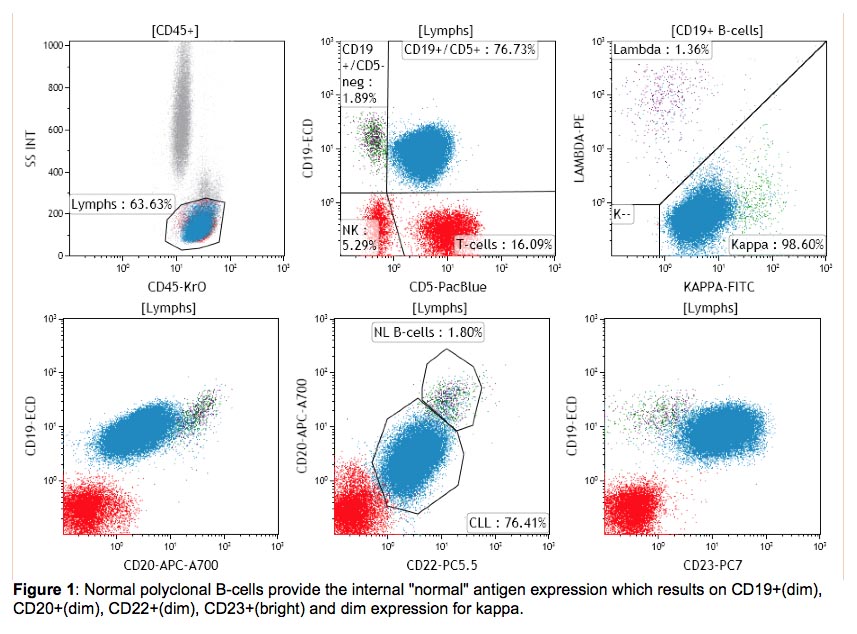
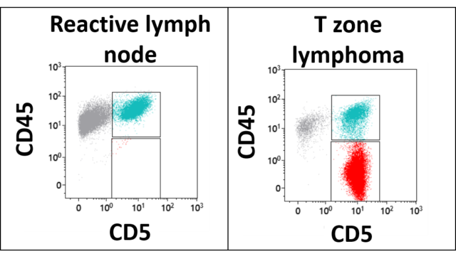
CD5 CD11c CD19 CD20 CD22 CD23 CD38 CD45 CD103 CD200 and kappa and lambda light chains. However flow cytometry results usually make certain lymphoma entities extremely likely and others very unlikely. CLL has a characteristic immunophenotype expressing CD5 CD19 dim CD20 dim CD22 CD23 bright CD43 dim CD45 dim to negative CD79b dim CD81 CD200 and dim monoclonal surface immunoglobulin.
The positive and negative predictive values of combined cytology and FCA in the patients with history of lymphoma andor abnormal imaging results were 92 and 89 respectively when compared with open brain tissue biopsy and 89 and 86 respectively when compared with clinical follow-up. It is used to detect abnormal hematolymphoid populations determine what cell surface markers they express and integrate immunophenotypic findings with morphologic and available clinical and laboratory data. When using fresh tissue for flow cytometric immunophenotyping the predominant populations are lymphoid.
Corticosteroids can induce apoptosis in lymphoma cells. Flow cytometry FC is usually recommended for the classification and staging of lymphomas in patients with organomegaly and atypical cells in effusions and blood after the exclusion of other possible diagnoses. This can mask the morphology and can even cause the tumor to vanish 31-33.
This flow cytometry test is used to diagnose leukemia or lymphoma. Flow cytometry is a laser-based technique used to detect and analyze the chemical and physical characteristics of cells or particles. Flow cytometric immunophenotyping of peripheral blood bone marrow and body fluids is performed using the following antibodies.
The blast markers CD34 and TdT are negative. FC may also have a place in the initial diagnostic investigation of aggressive lymphoma. Six cases had a benign immunophenotype.
Compensation matrix was calculated by using BD CompBead particles Becton Dickinson and the compensation setup tool in BD FACSDiva software. A pathologist often one specializing in the study of blood diseases andor blood cell cancers a hematopathologist will consider the results from the complete blood count CBC differential blood smear bone marrow findings and flow cytometry immunophenotyping as well as other tests in order to provide a diagnostic interpretation. Flow cytometry has a high specificity and can confirm the diagnosis of a lymphoma significantly faster than immunohistochemistry.
A laboratory report will typically. In lymph nodes and CSF samples flow cytometry can confirm a. CD10 CD38 CD43 CD71 and bcl-6 are also expressed.
Leukemia and lymphoma analysis by flow cytometry aids in identifying the tumor lineage which in most cases is identified as T cell B cell or myeloid. It is likely that this additional sensitivity of flow cytometry is a result of the low number of tumor cells available for diagnosis in CSF and bone marrow. It is most commonly used to evaluate bone marrow peripheral blood and other fluids in your body.
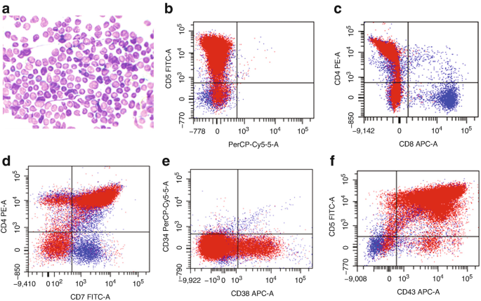
Case 1 showed several immunophenotypic deviations in addition to lack of SIg light chain expression. CD3 CD10 CD16 CD19 CD34 CD45 and kappa and lambda light chains. Case 1 have expression of B cellassociated antigens CD19 CD20 CD22 and CD79a by flow cytometry.
These results will explain if any abnormal cells are present and what types of cells they are as a part of your diagnosis. Phenotypic characteristics of BL are summarized in table below. This allows for rapid initiation of treatment in this highly.
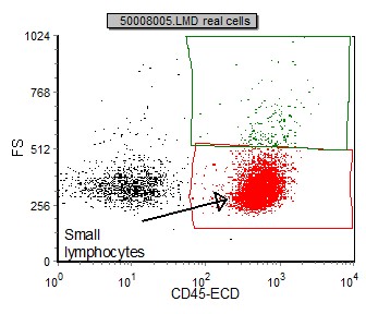
Not always strictly speaking not very often. These cells were in the subsequent anlysis. The gating dot plot below identifies a predominant CD45 bright FS small used cells.
This test generates a hematopathology report with a diagnosis and interpretation of findings. Tected by flow cytometry. These can be stratified as large and small lymphocytes CD45 positive.
The time between the biopsy and reporting the result turnaround time was significantly shorter for flow cytometry compared to immunohistochemistry median. Possible Additional Panels-B-cell Panel. Lineage identification can provide a confirmatory diagnosis or differential diagnosis prognosis and treatment options.
Flow cytometric analysis of lymphoma cells. Case 1 lack the expression of CD10 CD15 NG2 CD3 cCD3 MPO CD13 CD33 and CD7. FCM analysis was done on the FACSCantoII flow cytometer Becton Dickinson San Jose CA USA and the Kaluza software Beckman Coulter Brea CA USA.
Therefore flow cytometry is an important integral part of lymphoma diagnosis even in cases where it cannot give a definitive diagnosis. This test is usually done after atypical results are seen on a complete blood count or white blood cell WBC differential. Cytologic ex- amination was benign in 4 of these and suspicious for lymphoma in 2.

Selected Flow Cytometric Immunophenotyping Plots From Fine Needle Download Scientific Diagram
Flow Cytometry Analysis Of Ep On Apoptosis And Cell Cycle Progression Download Scientific Diagram
International Clinical Cytometry Society
Follicular Lymphoma Fl Flow Cytometry

Typical Data From A Two Color Flow Cytometry Experiment To Measure Cell Download Scientific Diagram

Diffuse Large B Cell Lymphoma Dlbcl Flow Cytometry

Flow Cytometry Demonstrating Cd10 Expression On Mycosis Download Scientific Diagram
Flow Cytometry Of Mature And Immature T Cell Lymphoma Springerlink
International Clinical Cytometry Society

Flow Cytometric Analysis Of Representative Tissue From The Inguinal Download Scientific Diagram

Flow Cytometry Market Size Trends And Forecast To 2027

International Clinical Cytometry Society
Principles Of Testing And Publications Clinical Hematopathology Laboratory

B Flow Cytometry On Peripheral Blood Revealed An Abnormal Population Download Scientific Diagram

Immunophenotype By Flow Cytometry Of The Peripheral Blood Showing Download Scientific Diagram

Flow Cytometric Immunophenotyping Performed On The Same Plasmablastic Download Scientific Diagram

Pb Flow Cytometric Analysis Download Table

Flow Cytometric Presentation Of A Large B Cell Lymphoma A Forward Download Scientific Diagram

A D Flow Cytometry Interpretation The Neoplastic Cells Display The Download Scientific Diagram